Fig. 3.
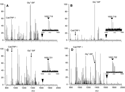
Direct tissue MALDI-FTMS spectra of freshly dissected commissural ganglia from (A) Petrolisthes cinctipes (infraorder Anomura), (B) Cancer magister (infraorder Brachyura), (C) Panulirus versicolor (infraorder Achelata) and (D) Callianassa californiensis (infraorder Thalassinidea). All spectra were measured using DHB as the matrix, with conditions optimized for m/z 1500. The inverted triangle shows the location of the m/z of the [M+H]+ ion for SYWKQCAFNAVSCFamide (disulfide bridge present between the cysteine residues; calculated m/z=1650.7192). The inserts show an expansion of the [M+H]+ peak region to show the measured mass and the isotopic distributions. Spectra were calibrated using known peptide peaks, including APSGFLGMRamide (CabTRP I) and GYRKPPFNGSIFamide (Gly1-SIF) at m/z 934.4927 and m/z 1381.7375, respectively.

