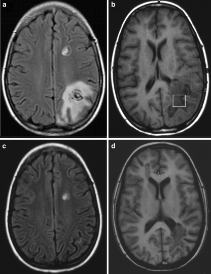
Fig. 1.
MRI of a 13-year-old child with Balo’s concentric sclerosis at (a,b) 2 months and (c,d) 3 months after onset of symptoms. a Axial FLAIR image demonstrating the concentric pattern of the two left-hemispheric lesions and b T1-weighted image depicting the VOI selected for proton MRS. c Axial FLAIR and d T1-weighted image showing regression of the left parieto-occipital lesion

