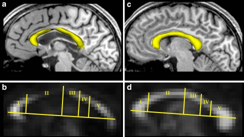
Fig. 5.
a,c Sagittal view of CC surface reconstructions (yellow) and b,d corresponding fractional anisotropy maps of the patient (a,b) and an age-matched control (c,d). The topographic scheme of the CC [26] identifies regions with fibers projecting into specific cortical areas: I prefrontal lobe, II premotor and supplementary motor cortex, III motor cortex, IV sensory cortex, V parietal, temporal, and occipital lobes

