
Figure 8. Comparison of myogenesis in 2D monolayers, tissue sheets and tissue networks.
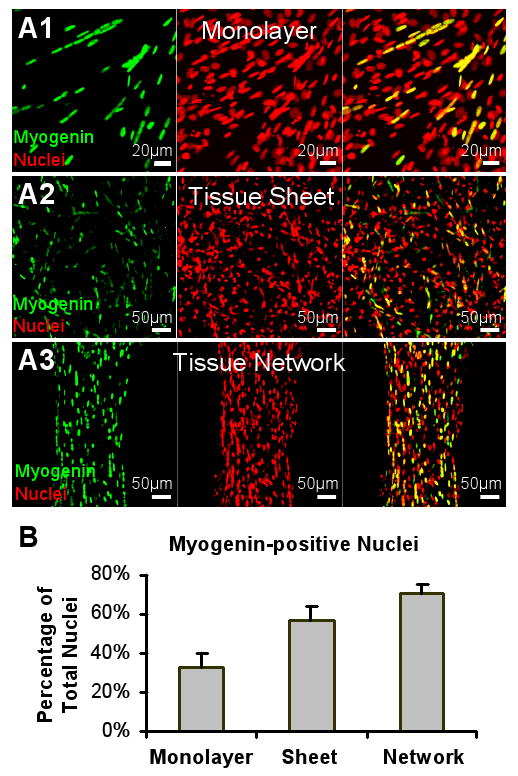
Representative images of myogenin-positive nuclei in a monolayer (A1), a non-porous tissue sheet (A2) and a porous tissue network (A3) after 4 days of differentiation. (B) The proportions of myogenin-positive nuclei significantly differ among the three groups.