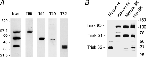
Figure 2. Western blot analysis of triadin isoforms.
A, the rat skeletal muscle isoforms, detected by an antibody in common region (Nter) or in specific C-terminal region of each isoform. B, the triadin isoforms expressed in different tissues (mouse heart, human skeletal muscle, mouse skeletal muscle, rat skeletal muscle) detected with an antibody against the common N-terminal part of all triadins, and therefore able to react with all the triadins and used to quantify their relative amount. H: heart; SK: skeletal muscle.

