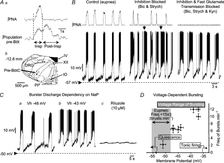
Figure 1. Example of discharges of burster neurones recorded intracellularly using patch pipettes from the ventral respiratory column.
Prior to patch recording, low impedance microelectrodes were used to map the respiratory column (A). Once mapped, patch pipettes were placed into this region and recordings made from inspiratory neurones during eupnoea (B). Note that membrane potential depolarized during neural expiration, with the discharge phase-locked with the phrenic bursts (∫ PNA; B). Following additions of bicuculline (Bicuc) and strychine (Strych) to the perfusate (Inhibition Blocked; B), neuronal bursting showed some uncoupling from the phrenic burst (arrowed). Endogenous bursting was revealed on blocking both inhibitory and excitatory (Kynurenic acid, Kyn) fast synaptic transmission – Inhibition and Fast Glutamate Transmission Blocked. Note the absence of any phrenic discharge but phasic firing of medullary burster. The location of the recording site of this neurone was in the pre-Bötzinger complex (Ab). There was a dependency of frequency of bursting upon membrane potential (Ca and b). Depolarization of neurones caused an increase in burst frequency (Ca and b and D). Note that burst frequency of this neurone did not match that seen during eupnoea even at the most depolarized levels. Bursting was eliminated following addition to the perfusate of 10 μm of riluzole, a blocker of persistent sodium channels (Cc). Small oscillations in membrane potential after riluzole most probably represent an incomplete blockade of currents through persistent sodium channels. Abbreviations: DRG, dorsal respiratory group; IO, inferior olive; Pre-BötC, pre-Bötzinger complex; pyr, pyramid; IV, fourth ventricle; XII, hypoglossal motor nucleus.

