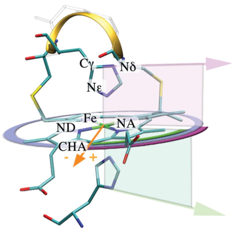
Figure 1.

Heme structure with definitions used in this paper. Structure of a typical c-type heme-binding motif is shown (1Z1N). The heme is ligated by two histidines and covalently attached to the protein via two thioether bonds made between two cysteine side chains and two heme vinyls. Ligation and attachment forms the classical c-type heme-binding motif Cys-Xaa-Xaa-Cys-His (CXXCH) illustrated as a ribbon. The orange vector (Fe-CHA) represents the heme basis vector (0°). The orientation of the histidine ligand (purple and green arrows), alpha is defined after the histidine vector (Cγ-Nδ) is projected onto the heme plane. The resulting projected ligand vectors unambiguously describe their orientation (purple and green arcs). The right hand rule (NA-NB-NC) defines the top (purple) and the bottom (green) side of the heme plane. Alpha of +45° and −45° is pointing towards the heme nitrogen NA and ND, respectively.
