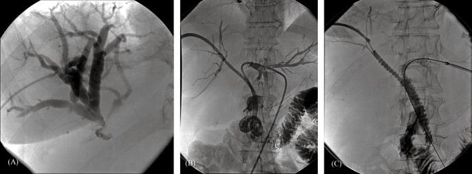
Figure 3.

A 71-year-old woman diagnosed with a cholangiocarcinoma. (A) Percutaneous transhepatic cholangiogram demonstrating dilated bile ducts of the right liver lobe with a stricture at the level of the hilum and no filling of the left bile duct or common bile duct. (B) Contrast medium injection after percutaneous transhepatic placement of drainage catheters through both the right and left liver lobes into the duodenum, showing a stricture at the level of the hilum involving the main left and right bile ducts. (C) Contrast filling of the bile ducts after placing Luminexx(tm) stents from the right and left sides. One of the stents protrudes into the duodenum
