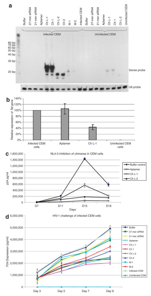
Figure 5. Dual inhibition on human immunodeficiency virus type 1 (HIV-1) infection mediated by small interfering RNA (siRNA) chimeras.
(a) Northern blots of infected CEM cells. Infected CEM cells were directly treated with siRNA and chimeras. The 27-chimera RNA is partially processed to a 21-mer siRNA after uptake into the CEM cells. Total RNAs were hybridized with a 21-mer 32P-labeled oligonucleotide probe. U6 RNA was used as an internal loading control. (b) Aptamer-mediated inhibition of expression of tat/rev in infected CEM cells. Cells were incubated with either the wild-type aptamer or Ch L-1 for 7 days before RNA extraction. Gene expressions for Tat/rev and glyceraldehyde-3-phosphate dehydrogenase were assayed using quantitative real-time PCR. The data shown represent the average of three replicate assays. (c) Chimera RNAs inhibit HIV infection. HIV-1 NL4-3 was incubated with the various RNAs at 37 °C for 1 hour. Subsequently, the treated virions were used for infecting CEM cells. The culture supernatant was collected at different time points (7, 11, 15, and 18 days) for p24 antigen analyses. The data shown represent the average of duplicate assays. (d) The siRNAs delivered by the chimera RNAs inhibit HIV-1 replication in previously infected CEM cells. Approximately 1.5 × 10⁴ infected CEM cells and 3.5 × 10⁴ uninfected CEM cells were incubated at 37 °C with the various RNAs at a final concentration of 400 nmol/l. The culture supernatant was collected at different time points (3, 5, 7, and 9 days) for p24 antigen analyses. The data shown represent the average of triplicate measurements of p24.

