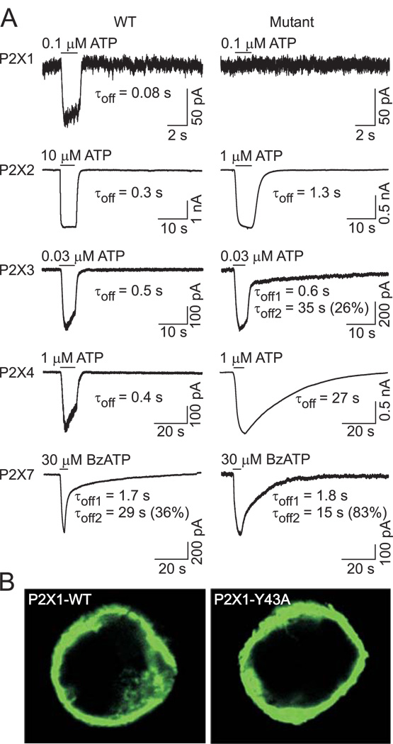
FIGURE 2. Effects of replacement of the conserved TM1 tyrosine residue with alanine on the expression and function of P2XRs.
(A) Example records of agonist-induced currents in cells expressing the wild type (WT, left) and α-position alanine mutants (right) of the P2X1R, P2X2R, P2X3R, P2X4R, and P2X7R. Currents were recorded from HEK293 cells using whole-cell patch clamp recording at a holding potential of −60 mV. Horizontal bars indicate the time of ATP or BzATP application. Numbers below the traces show deactivation time constant (τoff) values derived from mono-exponential fitting of current decay for the P2X1R, P2X2R, and P2X4R and bi-exponential fitting for the P2X3R and P2X7R; for the latter, the percentage of the slow component (τoff2) contribution to the current decay is also shown. (B) Expression pattern of the WT and non-functional P2X1R-Y43A mutant in HEK293 cells.

