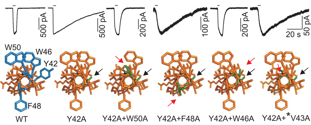
FIGURE 6. Replacement of ipsilateral but not contralateral TM1 aromatic residues with alanine restores P2X4R function.
Time-courses of currents from the P2X4R-WT and single- and double-point alanine mutants in response to brief 1µM ATP application (indicated by horizontal bars; top) and dependence of receptor deactivation on the position of the substituted residues in a helical model of the P2X4R (bottom). Helical models of P2X4R are viewed from the extracellular side with aromatic residues in the 42, 46, 48 an 50 positions (blue) and their alanine substitutions (green). Black arrows indicate alanine substitution of the conserved tyrosine, and red arrows indicate the second alanine substitution of another aromatic (Trp46, Trp50 and Phe48) residue or non-aromatic residue Val43 (*) present in the β position.

