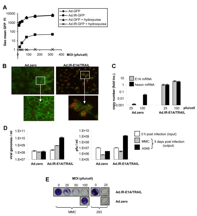
Fig. 1. Ad-replication in vitro.
A. GFP detection via flow cytometry in Ad.GFP and Ad.IR-GFP transduced MMC cells. (3 days post infection [p.i.]). Hydroxyurea was used to inhibit Ad-replication. B. Detection of hexon protein in Ad.zero and Ad.IR-E1A/TRAIL transduced cells (3 days p.i.; MOI 100 pfu/cell; positive cells show red nuclear staining; anti-E-cadherin-FITC was used as a cell surface marker). Representative pictures are shown, magnification 20× (upper panel); 40× (lower panel). C. Relative quantification of E1A and hexon mRNA expression in Ad.zero and Ad.IR-E1A/TRAIL infected MMC cells using real-time RT PCR (7 days p.i.). y=1 for Ad.IR-E1A/TRAIL infected cells (MOI 25 pfu/cell). D. Absolute quantification of viral genomes and plaque forming units (pfu) in Ad.zero and Ad.IR-E1A/TRAIL infected MMC and A549 cells (0h and 5 days p.i.). E. CPE formation in MMC and 293 cells after Ad.zero and Ad.IR-E1A/TRAIL infection (5 days p.i.).; D,E,: Bars indicate the mean values and SD of experiments performed in duplicates.

