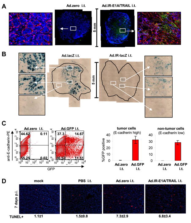
Fig. 3. Ad-vector function in vivo.
For all experiments 5×10⁵ MMC cells were injected subcutaneously into neu-tg mice and 1×10⁹ pfu Ad-vector was injected once intratumorally when tumors reached a size of 3–4 mm diameter. A. Hexon expression in MMC tumors 3 days after injection with Ad.zero or Ad.IR-E1A/TRAIL (n=3 animals/group). Tumors were harvested, fixed, sectioned and stained for hexon (green), laminin (red) and nuclei (blue). Representative whole tumor sections (middle panels) and 20× magnifications (side panels) are shown. Laminin staining is only shown in the 20× magnifications. B. LacZ expression in MMC tumors 3 days after injection with Ad.lacZ or Ad.IR-lacZ (n=3 animals/group). Tumors were extracted, fixed, sectioned and stained for lacZ. Representative whole tumor sections (middle panels) and 20× magnifications (side panels) are shown. C. GFP expression in MMC tumors 24 h after injection with Ad.zero or Ad.GFP (n=3 animals/group). Tumors were collected, dispersed into single cells and analyzed for E-cadherin and GFP expression via flow cytometry. Percentages of GFP−/+ MMC cells (E-cadherin high) and GFP−/+ non-MMC cells (E-cadherin low) were determined. Left panel: Representative flow charts are shown. Right panel: Bars indicate mean and SD. D. Detection of apoptotic cells in MMC tumors 5 days after intratumoral injection with Ad.zero or Ad.IR-E1A/TRAIL (n=3 animals/group). Tumors were extracted, fixed, sectioned and stained for TUNEL positive cells (red) and nuclei (blue). Representative pictures are shown (Magnification 20×). Mean number of TUNEL positive cells ± one SD are indicated.

