
Fig. 2.
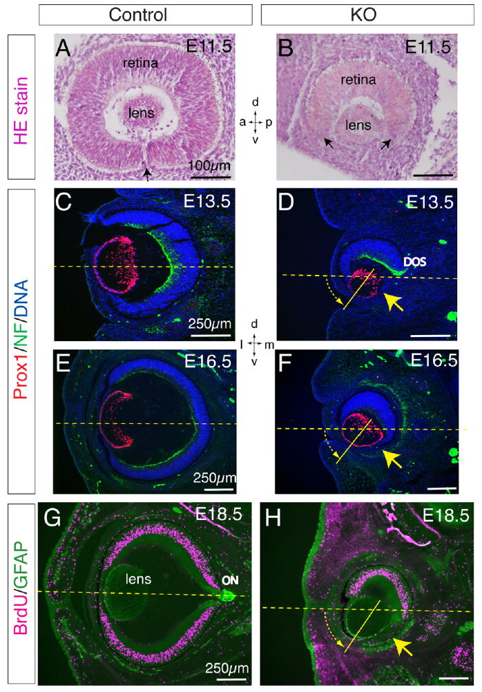
Histological analyses of retinal defects and lens rotation in Lrp6-knockout (KO) embryos. A,B: Hematoxylin & eosin staining of the frontal eye sections of the normal control and Lrp6-KO embryos at embryonic day (E) 11.5. Arrow in A indicates the closing site of the ventral retina. Arrows in B indicate the retinal coloboma, a big gap between the ventral ends of the retina (refer to arrows in Fig. 1B). C–H: Coronal head sections through mid eye regions of the control (C,E,G) and Lrp6-KO (D,F,H) embryos demonstrate the complete absence of the neuroretina and retinal pigment epithelium (RPE) in the ventral-middle eye of Lrp6-KO (yellow arrows; refer to arrows in Fig. 1D,F). C–F: Double immunolabeling for Prox1 (red, lens cells) and neurofilament protein (NF, green, retinal ganglion axons) at E13.5 (C,D) and E16.5 (E,F). Only the dorsal optic stalk (DOS; green in D) immunolabeled by neurofilament is observed in the mutant eye. Nuclei were counterstained by 4′,6-diamidine-2-phenylidole-dihydrochloride (DAPI). G,H: Double immunolabeling for bromodeoxyuridine (BrdU) incorporation (red, proliferating cells) and glial fibrillary acidic protein (GFAP; green, mesenchymal and lens epithelial cells) at E18.5. A group of putative mesenchymal cells immunolabeled by GFAP is absent in the mutant eyelids (H). The vitreous body (fills the space behind the lens) is also defective in the mutant eyes (D,F,H). ON, optic nerve. Note the ventral 45° rotation approximately of the lens in the mutant eye (shown with yellow dashed arrow). Dashed lines in C–H indicate the horizontal midline of the eye. Axes: d, dorsal; v, ventral; a, anterior; p, posterior; l, lateral; m, medial.