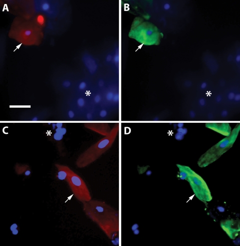
Figure 1.
Identification of cytotrophoblast cells in cervical samples by immunofluorescence microscopy. Cervical cells were labeled with antibodies against HLA-G (A, C; red) and cytokeratin 7 (B, D; green), as well as with DAPI to stain nuclei (shown in all panels; blue). Cells were identified as cytotrophoblasts by their labeling for both HLA-G and cytokeratin (indicated with arrows). Resident cervical cells appear as DAPI stained cells that were not labeled by either antibody (indicated by asterisks). Bar, 50 µm.

