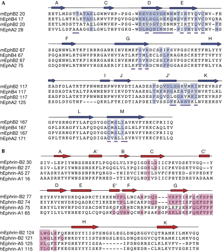
Figure 2.
Structure-based sequence alignment of Eph receptors and ephrins. Secondary structure elements are shown above the sequences. The residues forming the high-affinity ligand–receptor interfaces are highlighted in blue and red. (A) Sequence alignment of Eph receptors the structure of which, bound to their ligands, has been reported. The residues forming the EphA2 homodimer interface observed in the unligated structure have a magenta underscore. (B) Sequence alignment of ephrins the structure of which, bound to their receptors, has been reported.

