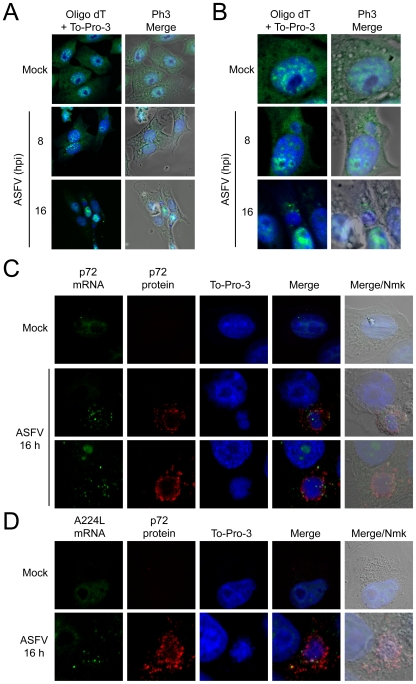
Figure 7. Localization of mRNAs in ASFV-infected Vero cells.
Vero cells were seeded on glass coverslips and mock infected or infected with 5 pfu/cell of ASFV. At 8 and 16 hpi cells were fixed and permeabilized. Then, in situ hybridization with fluorescein labeled probes was carried out for each post-infection time. A) Distribution of polyadenylated mRNAs bulk by using oligo d(T) fluoresecein labelled probe. To-Pro 3 was used to stain cell nuclei and ASFV factories. B) Detail of mock infected and ASFV-infected cells, showing the polyadenylated mRNAs surrounding the viral factories. In situ hybridization with fluorescein labeled p72 (C) and A224L (D) probes. To-Pro 3 and p72 antibody were used to detect cell nuclei and ASFV factories. Cells were visualized by confocal microscopy and the cell outline was defined by phase contrast microscopy. Images were processed with Huygens 3.0 software.

