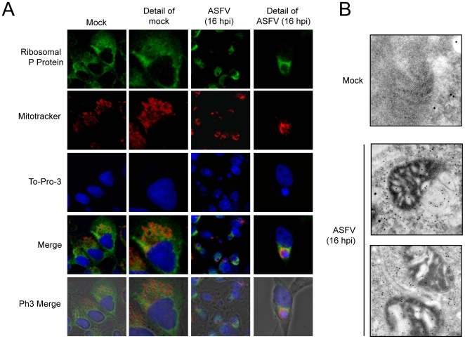
Figure 9. P ribosomal protein and mitochondrial network co-localize surrounding ASFV factories.
A) Vero cells were seeded on glass coverslips and infected with 5 pfu/cell of ASFV. For mitochondrial staining, cells were incubated at 15 hpi with 2 µM MitoTracker red CMH2-Ros for 45 min and then permeabilized and fixed. Ribosomal P protein was detected by indirect immunofluorescence and cell nuclei and viral factories were stained with To-Pro-3. Cells were visualized by confocal microscopy and the cell outline was defined by phase contrast microscopy. Images were obtained under restricted conditions and processed with Huygens 3.0 software. B) Detection by electron microscopy of ribosomes in mitochondria-containing areas. Cells were mock infected or infected with 5 pfu/cell of ASFV and processed for electron microscopy at 16 hpi. Ribosomes were detected using a specific monoclonal antibody against P ribosomal protein.

