
Fig. 1.
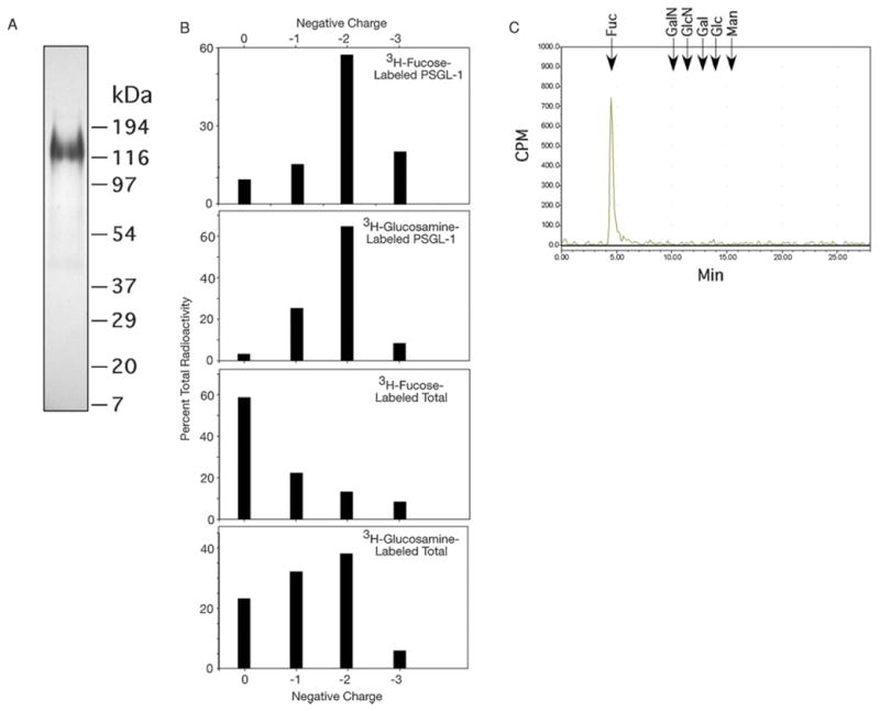
(A) SDS–PAGE and autoradiography of 3H-fucose-labeled PSGL-1 immunoaffinity purified from WEHI-3 cells. (B) The β-eliminated O-glycans from radiolabeled PSGL-1 and total cellular glycoproteins were separated and eluted to collect glycans of neutral structure (0 charge), 1 sialic acid (−1 charge), 2 sialic acids (−2 charge), or >2 sialic acids (−3 charge). The distribution of total radioactivity as a percentage from individual samples in the four major fractions is shown. (C) Monosaccharide analysis of O-glycans from 3H-fucose-labeled WEHI-3 cells. Elution positions of standards are indicated. Fuc, fucose; GalN, galactosamine; GlcN, glucosamine; Gal, galactose; Glc, glucose; Man, mannose.