Fig. 1.
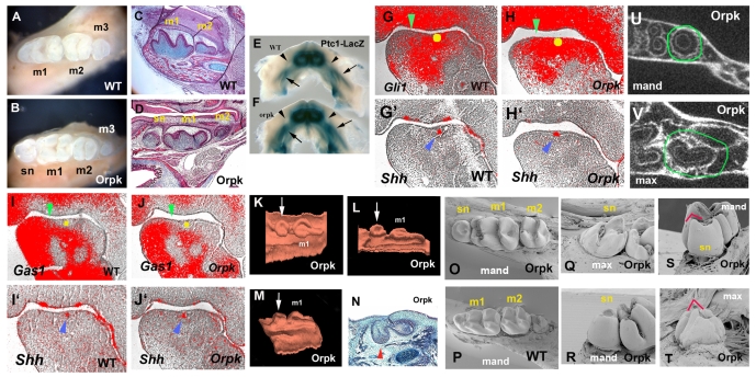
Lower molar tooth phenotypes and Shh signalling in Tg737orpk homozygous mutant mice. (A-D) Whole teeth (A,B) and sagittal sections (C,D) show that supernumerary teeth develop mesial to the first molars (sn in B,D) in mutants. (E,F) Increase of Ptch1-lacZ staining in the mandible of Tg737orpk mutants with Ptch1-lacZ allele at E13.5. Ptch1-lacZ staining was expanded into the diastema region (arrowhead). Arrows indicate molar germs. (G-J) Radioactive in situ hybridisation on sagittal sections showing Gli1 (G,H), Gas1 (I,J) and Shh (G',H',I',J') expression in mandibles of embryo heads at E12.5. (G,H,I,J) Yellow circles represent positions of Shh expression in adjacent sections (blue arrowheads in G',H',I',J'). Gli1 expression (H) was expanded into the diastema and Gas1 (J) was downregulated in the diastema of Tg737orpk mutants (green arrowheads). (K-N) Sagittal sections (N) and three-dimensional reconstructions (K-M) of the dental epithelium of the mandible molar region at E15.5 (N) and E16.5 (K-M) in Tg737orpk mutants. The supernumerary tooth buds (arrows in K-M) were observed in the position of the mesial swellings (arrowhead in N). (O-T) SEM analysis shows that the lingual cusp of maxillary supernumerary tooth (red line in T) is more prominent in comparison with that of mandibular supernumerary tooth (red line in S). (U,V) Horizontal micro-CT sections of Tg737orpk show that maxillary supernumerary teeth have concave roots (circle in V) and in mandibles show round roots (circle in U). m1, first molar; m2, second molar; m3, third molar; mand, mandible jaw; max, maxillary jaw.

