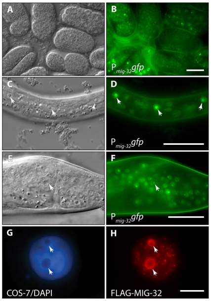
Fig. 3.
mig-32 is broadly expressed, localized to nuclei, and concentrated in nucleoli. Differential interference contrast (A,C,E) and epifluorescence (B,D,F) images of transgenic C. elegans. (A,B) Mixed-stage embryos. (C,D) L1-stage larva showing the lateral hypodermal cells. Arrowheads highlight nucleoli within hypodermal nuclei. (E,F) L4-stage male tail. Arrowhead highlights a nucleolus within a neuronal nucleus. (G,H) COS-7 cell transfected with FLAG-epitope-tagged C. elegans MIG-32, stained with (G) DAPI to visualize DNA and (H) anti-FLAG antibody. Arrowheads highlight nucleoli in the COS cell nucleus. In C-F, anterior is to the left and ventral down. Scale bars: 10 μm.

