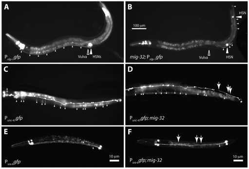
Fig. 5.
mig-32 mutants have defects in neuronal migration and process extension. Images of wild-type and mig-32 mutant animals carrying gfp reporters. (A) An otherwise wild-type animal expressing Pnlp-1gfp in the HSN neurons. HSN cell bodies are indicated with a large white arrowhead. The position of the vulva is indicated. Small white arrowheads highlight the axons of the two HSN neurons, which proceed along the ventral body wall to the head. (B)A mig-32 mutant expressing Pnlp-1gfp. Large white arrowheads indicate the HSN cell bodies and small arrowheads highlight the HSN axons. (C) Ventral view of a wild-type animal expressing Punc-47gfp in the VD neurons. Small white arrowheads indicate lateral commissures. (D) Ventral view of a mig-32 mutant expressing Punc-47gfp in the VD neurons. Small white arrowheads indicate lateral commissures. Large arrows indicate three commissures on the wrong side. (E) A wild-type animal expressing Psra-6gfp in the PVQL and PVQR neurons. A small white arrowhead indicates the normal separation of the PVQ axons into the right and left sides of the ventral nerve cord. (F)A mig-32 mutant expressing Psra-6gfp in the PVQ neurons. Arrows indicate inappropriate crossing by the PVQ neuronal axons. The scale bar in B applies to A-D, which show adult animals. E and F show L1-stage larvae. Anterior is to the left in all images; ventral is down in A and B, up in C and D, and slightly rotated in E and F.

