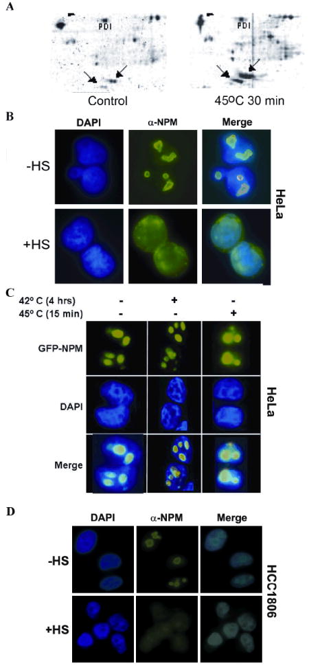
Figure 1. Nucleophosmin redistribution following heat shock.

(A) Proteins were crosslinked to DNA by cisplatin from control and heat shocked HeLaS3 cells. Two forms of NPM, B23.1 and B23.2, are denoted by arrows. (B) HeLaS3 cells were incubated at 45°C for 45’, fixed and stained with antibodies recognizing NPM. DNA was visualized with Hoechst dye and NPM with FITC filters. (C) Cells expressing a GFP-NPM fusion protein and counter stained with DAPI. (D) Redistribution of NPM following heat shock in HCC1806 cells.
