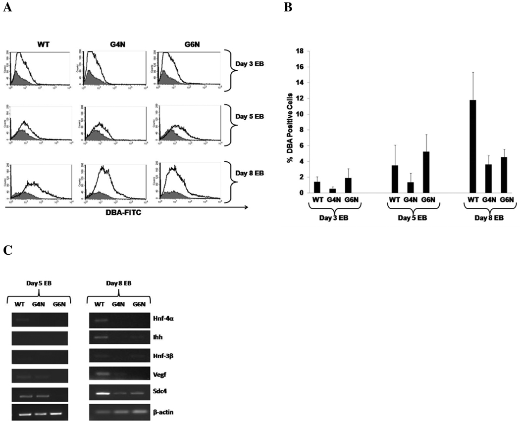
Figure 2. Diminished Visceral Endoderm Formation and Gene Expression in G4N and G6N EBs.
Cells from day 3, day 5 and day 6 EBs were stained with FITC-conjugated DBA and positive bound cells were analyzed by FACS analysis. (Representative experiment) (A). The average of four independent experiments are displayed (B). DBA positively bound cells were sorted from day 5 and day8 WT, G4N and G6N EBs. Total cellular RNA was isolated and reverse transcription was performed using 2.0 µg as template. Hnf-4α, Ihh, Hnf-3β, Vegf, Sdc4 and β-actin were amplified with 28 cycles of PCR. Products were visualized on agarose gel stained with ethidium bromide (C).

