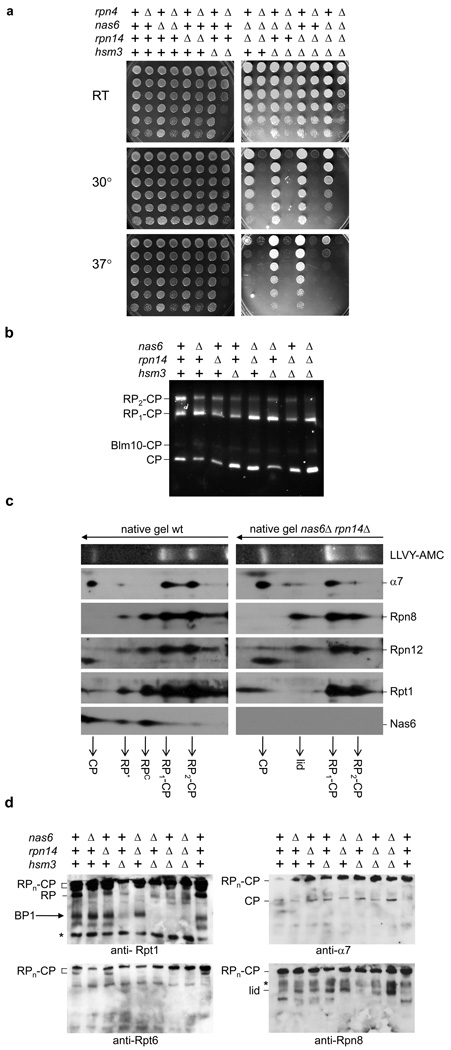
Figure 2. Phenotypic analysis of nas6Δ, hsm3Δ, and rpn14Δ mutants.
a, Strains with indicated genes deleted were spotted on plates in four-fold dilutions and grown at the indicated temperature.
b, Cell lysates (75µg) were resolved on native gels. Gels were stained for LLVY-AMC hydrolytic activity.
c, Cell lysates of wild-type or nas6Δ rpn14Δ strains were resolved by two-dimensional native-SDS/PAGE gel electrophoresis followed by immunoblotting.
d, Cell lysates were resolved on 5.25% native gels and immunoblotted. BP1 assignment based on ref. 2. *, background band.

