
Fig. 3. Combinatorial treatment with 17-AAG and Trastuzumab induces enhanced ubiquitinylation and lysosomal degradation of ErbB2.
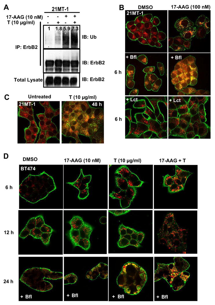
A. 21MT-1 cells were treated with the indicated concentrations of 17-AAG or Trastuzumab or the combination for 6h. Following treatment, the cells lysed and subjected to anti-ErbB2 IP according to previously described method (20). After SDS-PAGE/WB the membrane filter was probed for ubiquitin. The membrane filter was stripped and probed for ErbB2. 50 μg aliquots of total lysate were also similarly analyzed. B. 17-AAG induces ErbB2 accumulation in the lysosome; 21MT-1 cells grown on coverslips were left untreated (top row) or pretreated with 100 nM of the lysosomal inhibitor bafilomycin A1 (middle row) or 10 μM proteasomal inhibitor, lactacystin (bottom row) for 1 h. The cells were then either untreated (left column) or treated with 100 nM 17-AAG (right column). Following treatment, the coverslips were fixed in 4% paraformaldehyde (PFA) and immunostained for ErbB2 (green) and LAMP-1 (red) as detailed in the methods section. Shown here are the Confocal Immunofluorescence microscopy images. C. Trastuzumab-treatment also induces lysosomal localization of ErbB2; 21MT-1 cells, grown on coverslips, were untreated or treated with 10 μg/ml Trastuzumab for 48 h, before fixation and immunostained for ErbB2 (green) and LAMP-1 (red). D. BT-474 cells, grown on coverslips were treated with the indicated concentrations of single drugs or combination for the indicated time periods, with or without Bafilomycin (10 nM). The coverslips, after treatment, were processed as described in the legend for Fig. 3A. Shown here is ErbB2 staining in green and LAMP-1 in red; colocalized regions are seen in yellow.