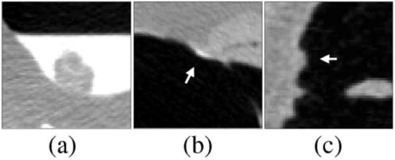
Fig. 10.

Comparison of typical FP and false-negative detections of the nCAD and ftCAD schemes. (a) The nCAD scheme missed this polyp covered by tagged fluid, whereas the ftCAD scheme detected the polyp correctly. (b) The nCAD scheme detected this stool (indicated by arrow) incorrectly as a flat lesion based upon its shape. The ftCAD scheme identified the lesion correctly as an FP based on the fecal tagging. (c) An example of untagged stool (indicated by arrow) that was detected incorrectly as a polyp by both CAD schemes.
