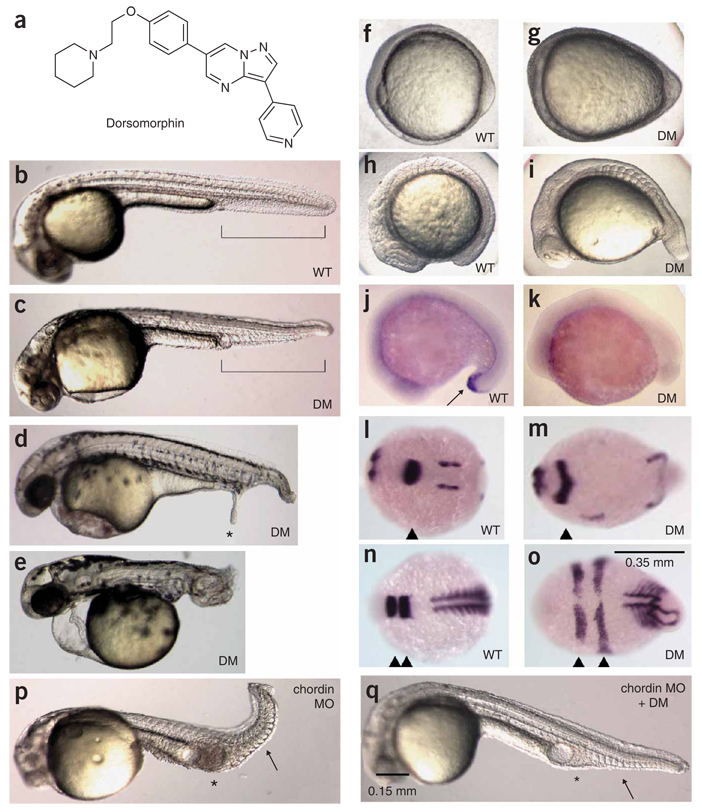
Figure 1.
Dorsomorphin induces dorsalization in zebrafish embryos. (a) Structure of dorsomorphin. (b) Vehicle-treated WT zebrafish embryo 36 h.p.f. Ventral tail fin is highlighted in brackets. (c) Zebrafish embryo treated with 10 µM dorsomorphin (DM) at 6–8 h.p.f. and photographed at 36 h.p.f. (d) Zebrafish embryos treated with 10 µM dorsomorphin at 6 h.p.f. occasionally develop ectopic tails (*) at 48 h.p.f. (e) Embryos treated with 10 µM dorsomorphin at 1–2 h.p.f. show severe dorsalization at 48 h.p.f. Embryos b–e are shown on lateral view. (f,g) Bud stage embryos left untreated (f) or treated at 2 h.p.f. with dorsomorphin (g) are shown on ventral view. (h,i) 10-somite-stage embryos left untreated (h) or treated at 2 h.p.f. with dorsomorphin (i) are shown on lateral view. (j,k) In situ hybridization of 18-somite stage WT embryos (lateral view) reveals typical expression of the ventral marker eve1 (arrow) in the distal tail (j), whereas embryos treated with dorsomorphin (10 µM) at 1 h.p.f. do not (k). (l–o) In situ hybridization of 6-somite-stage (12 h.p.f.) embryos (dorsal view, anterior left). The mid-hindbrain boundary is marked by pax2a (l and m, arrowheads). Rhombomeres 3 and 5 are marked by egr2b (krox20) (n and o, arrowheads), and somitic mesoderm is marked by myod (n and o). (p,q) Embryos injected with 1 ng chordin morpholino were treated with vehicle (p) or 20 µM dorsomorphin at 2 h.p.f. (q) and photographed 36 h.p.f. Dorsomorphin treatment normalizes expanded intermediate cell mass (*) and ventral tail fin structures (arrow).

