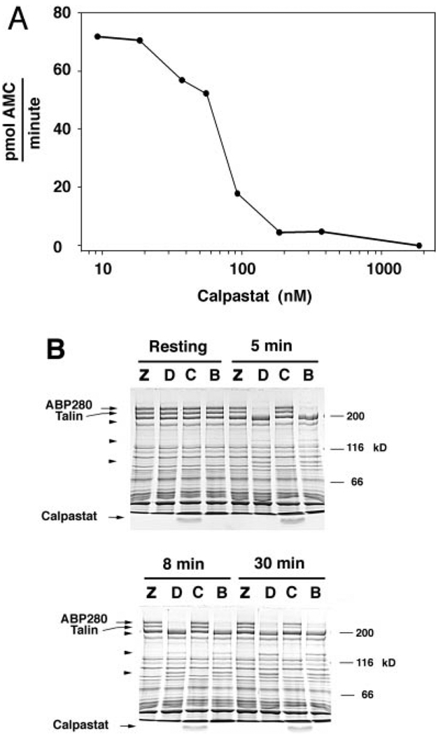
FIG. 2. Calpastat inhibits calpain in vitro.
A, the dose-response curve of calpastat for inhibition of μ-calpain cleavage of the substrate suc-LLVY-AMC, assayed as described (see “Experimental Procedures”). The IC50 of calpastat for inhibition of μ-calpain is 70 nm. The calpastat-Ala mutant peptide has no inhibitory activity in this assay. B, the cell-penetrating calpastatin peptide inhibits calpain in vivo. Platelets were preincubated with ZLLYCHN2 (Z), Me2SO vehicle (D), calpastat (C), or HEPES vehicle for calpastat (B), and treated with the ionophore A23187 for 0, 5, 8, or 30 min. Platelet proteins were analyzed by SDS-PAGE, as described (see “Experimental Procedures”). Arrowheads designate the 135- and 93-kDa calpain breakdown products of ABP280 and the 190-kDa calpain breakdown product of talin. The 49-kDa calpain breakdown product of talin is not visualized in this gel system. Free calpastat is visualized at the bottom of lanes corresponding to conditions where the peptide was added.

