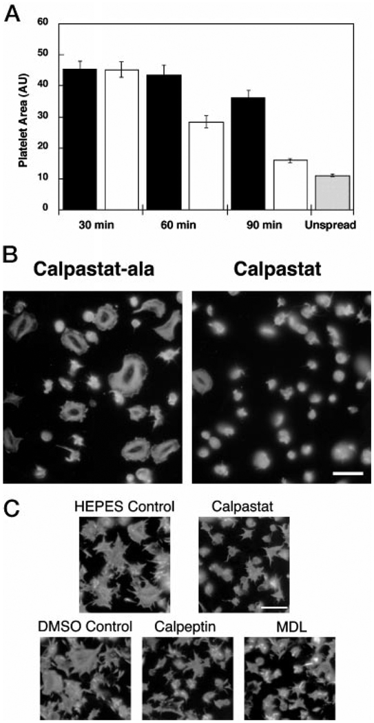
FIG. 5. Spreading of platelets on glass is calpain-dependent.
A, calpastat inhibits platelet spreading, dependent upon the calpastatin consensus sequence, and requires a pre-incubation period of between 30 and 60 min. Platelets were preincubated with calpastat-Ala (black bar) or calpastat (white bar) peptide (100 µm) for 30, 60, or 90 min and then spread for 20 min on glass coverslips. The coverslips were fixed, stained with Oregon Green-phalloidin, and photographed by fluorescence microscopy. The cell areas of the spread platelets were measured by computerized image analysis (NIH Image 1.61). One area unit (AU) is 0.625 µm2. Although calpastat-Ala demonstrated a 20% nonspecific inhibition of spreading relative to a vehicle control at 90 min of preincubation (data not shown), this inhibition was 4-fold less than the 85% inhibition observed for calpastat, compared with the vehicle control. Unspread platelets (gray bar) were obtained by treating platelets with 3% Me2SO, which completely blocks spreading, and allowing them to adhere to glass coverslips for 20 min as above. B, calpastat inhibits platelet actin remodeling during spreading, dependent upon the calpastatin consensus sequence. Platelets were incubated with peptide for 90 min and then spread on glass for 20 min. The coverslips were processed, stained with Oregon Green-phalloidin, and photographed by fluorescence microscopy. The size bar is 10 µm. C, similar to calpastat, MDL and calpeptin inhibit platelet actin remodeling during spreading. Calpastat (100 µm), MDL (400 µm), and calpeptin (300 µm) were used at their respective IC60 concentrations for spreading inhibition. Preincubation times were 90 min for calpastat and HEPES vehicle control and 10 min for MDL, calpeptin, and the Me2SO vehicle control. NH4Cl (10 µm) treatment for 10 min had no detectable effect on spreading (data not shown). The coverslips were processed, stained with Oregon Green-phalloidin, and photographed by fluorescence microscopy. The size bar is 10 µm. The IC60 concentrations of inhibitors were chosen to demonstrate the morphologies of the actin cytoskeleton for calpastat and peptidyl calpain inhibitor-treated platelets, under conditions of partial and equal spreading.

