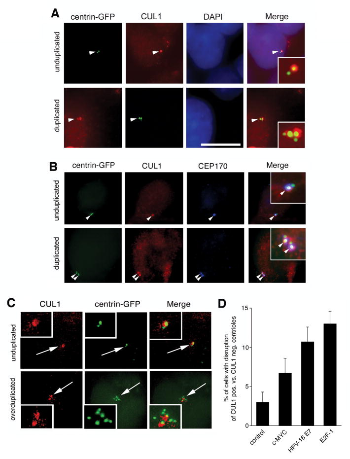
Figure 1. CUL1-positive maternal centrioles serve as assembly platforms for oncogene-induced centriole overduplication.
(A) Immunofluorescence microscopic analysis for CUL1 using U-2 OS cells stably expressing centrin-GFP (U-2 OS/centrin-GFP). Arrowheads indicate centrioles shown in inserts. Nuclei stained with DAPI. Scale bar indicates 10 μm.
(B) Co-immunofluorescence microscopic analysis of U-2 OS/centrin-GFP cells for CUL1 and CEP170, a marker for mature maternal centrioles. Arrowheads indicate centrioles with co-localization of CUL1 and CEP170 (see also inserts).
(C) Immunofluorescence microscopic analysis of U-2 OS/centrin-GFP cells for CUL1 following overexpression of E2F-1. Note overduplication of centrioles in the presence of only two CUL1-positive centrioles (bottom panels).
(D) Quantification of the proportion of U-2 OS/centrin-GFP cells with centriole overduplication in the presence of one or two CUL1-positive centrioles after overexpression of c-MYC, HPV-16 E7 or E2F-1. Arrows point to centrioles shown in inserts. Mean and standard error of three independent experiments with at least 100 cells counted per experiment are shown.

