
Figure 4. PLK4 is degraded by the proteasome.
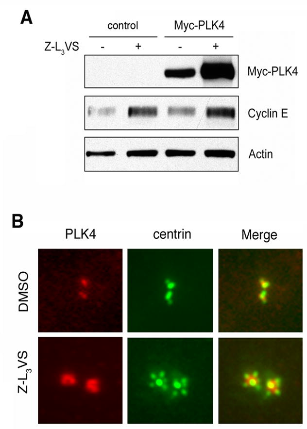
(A) Immunoblot analysis of whole cell extracts from U-2 OS/centrin-GFP cells transiently transfected with either empty vector (control) or PLK4 and treated with 1 μM of the proteasome inhibitor Z-L3VS or 0.1% DMSO at 24 h after transfection for an additional 24 h.
(B) Immunofluorescence microscopic analysis or U-2 OS/centrin-GFP cells treated with 0.1% DMSO or 1 μM Z-L3VS for 48 h and stained for PLK4. Note centriole multiplication together with an excessive amount of PLK4 at maternal centrioles in Z-L3VS-treated cells