
Figure 1. Cortical interneuron subgroup identity is determined within the MGE.
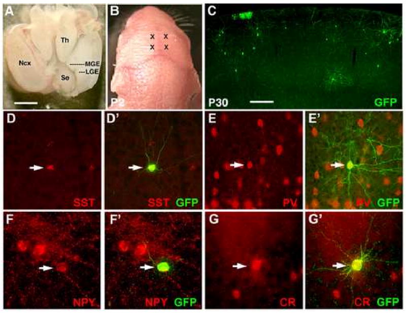
(A) The brain of an E13.5 embryo as viewed from above. Rostral is to the panel bottom. To isolate the MGE, the neocortex (Ncx) was dissected away as shown for the right hemisphere. The sulcus between the MGE and LGE is extended slightly, and a cut made along the medial-lateral axis at the level of the thalamus (Th). The MGE can then be separated free from the remainder of the brain. (B) Two injection sites per hemisphere were made into each neonate (marked by X's). (C) By P30, transplanted GFP+ cells are found throughout the cortex, of which the majority has adopted cortical interneuron morphologies. (D-G) Transplanted cells co-express interneuron subgroup markers including somatostatin (D-D′), parvalbumin (E-E′), neuropeptide Y (F-F′), and calretinin (G-G′). Scale bar in (A) is 1mm, in (C) is 200μm.