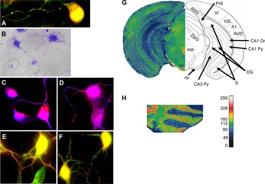
FIGURE 1.
MHC-I and Ly49 distribution and expression in cultured cortical neurons and their mRNA expression in the adult mouse brain. A, Codistribution of Abs specific for MHC I (red) and NK receptor Ly49G2 (green). In 1-day neurons MHC-I and Ly 49 colocalize (yellow) in somas and in some axonal regions. MHC-I primarily stains neurites in a clear punctate manner while Ly49 stains axons and dendrites. B, Distribution of SIINFEKL-FITC (blue; overlaid on the transmitted light image of the neurons in gray) in 1-day cell bodies and axons. C, MHC-I (blue) and synapsin I (red) show colocalization in cell bodies (purple) whereas in the axons they show alternate punctate distribution. D, MHC-I (blue) and Shank (red) also show colocalization in the cell bodies (purple) and alternating punctate distribution in axons. E, Ly49 (green) and synapsin-I (red) show colocalization (yellow) in cell bodies and in neurites. F, Ly49 (green) and Shank (red) show co localization (yellow) in cell bodies and alternating punctate distribution in axons. G, Cy3-cytidine 5′-triphosphate-labeled specific Ly49a probe was used to reveal its mRNA expression in adult mouse brain by in situ hybridization (see side bar next to H for intensities in pseudocolor.) Ly49a was expressed in the cerebral sensory cortex (VI, primary visual cortex; V2L, secondary visual cortex; A1, primary auditory cortex; AUD, secondary visual cortex), the hippocampus (CA1 Py, CA1 pyramidal cell layer; CA1 Or, CA1 oriens layer; CA3 Py, CA1 pyramidal cell layer; DG, dentate gyrus) and some of the pathways connecting the cortex and the hippocampus (RSG, retrosplenial cortex; PrS, presubiculum; S, subiculum). In the diencephalon expression was observed in areas containing en passent (scp and cp) or terminating (DpG) cerebellar axons, and also axons from other origins. H, Ly49a mRNA expression in the cerebellum was high in the Purkinje cell layer and moderate in the molecular and granule cell layer.

