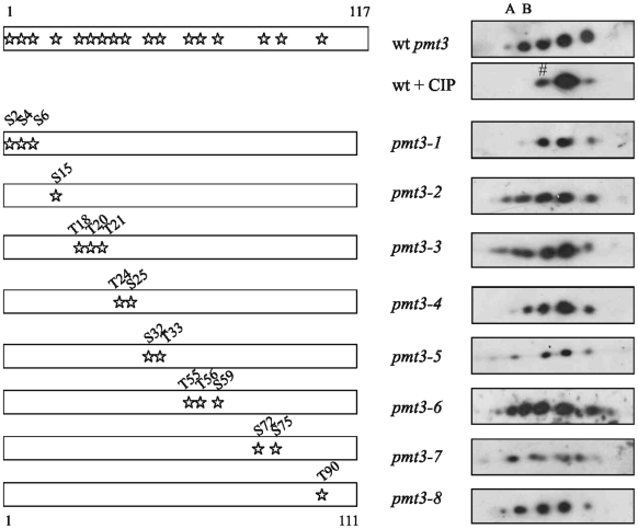
Figure 2. Position of serine and threonine residues and effects of mutations in SUMO/Pmt3.
Position of serine and threonine residues in SUMO/Pmt3 are indicated by stars. Left hand panel: sites of mutations in pmt3 mutants. Right hand panel: Western analysis with anti-SUMO antisera of 2D PAGE (1st dimension pH range 3–6, 2nd dimension 12.5% acrylamide) of extracts from wild type (wt), wild type+CIP and pmt3 mutants. A,B, forms not observed after CIP treatment, # possible acetylated form.

