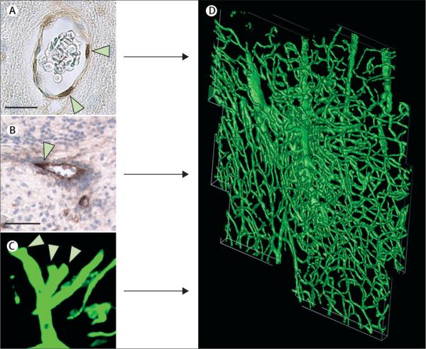
Figure 2. Stroke induces angiogenesis within the ischaemic boundary.
Immunostaining with antibodies against BrdU shows proliferative endothelial cells of cerebral blood vessels (A, arrowheads). Sprouting cerebral vessels are detected with immunoreactive von Willebrand factor (B, arrowhead) and shown on three-dimensional images obtained from confocal microscopy (C, arrowheads). Proliferating endothelial cells and sprouting vessels contribute to angiogenesis seen at the ischaemic boundary region (D). D is a three-dimensional image of angiogenesis at the cortical ischaemic boundary of a rat 14 days after stroke. Bars are 10 μm (A) and 50 μm (B). BrdU=5-bromo-2'-deoxyuridine.

