
FIG. 3.
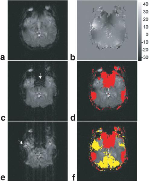
Type 1 and Type 2 artifacts in partial-Fourier gradient-echo EPI of human brain, simulated from the acquired full Fourier EPI data (through data truncation). a: The full Fourier EPI image of matrix size 96 • 96. b: The ky displacement map calculated from full Fourier EPI data using the k-space energy spectrum analysis. c: The partial Fourier EPI image reconstructed with Cuppen's algorithm and the full knowledge of image background phase (that is available from full Fourier EPI data). d: Image pixels susceptible to Type 1 artifact (shown in red) using the information from the ky displacement map (b). e: The partial Fourier EPI image reconstructed with Cuppen's algorithm and low-resolution estimation of image background phase (from the central ky band 96 • 12). f: Image pixels susceptible to Type 1 (shown in red) and Type 2 (shown in yellow) artifacts using the information from the ky displacement map (b).