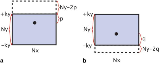
FIG. 5.

The k-space coverage in the multischeme reconstruction algorithm. a: When the k-space energy peak is closer to the most positive acquired ky lines, the reconstructed k-space coverage should be extended along the positive ky domain by Ny–2p lines, where p is the distance between the energy peak and the most positive acquired ky line. b: When the k-space energy peak is closer to the most negative acquired ky lines, the reconstructed k-space coverage should be extended along the negative ky domain by Ny–2q lines, where q is the distance between the energy peak and the most negative acquired ky line. [Color figure can be viewed in the online issue, which is available at http://www.interscience.wiley.com.]
