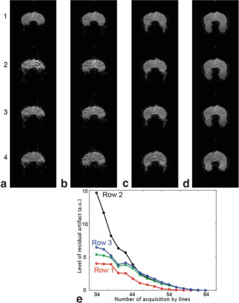
FIG. 6.

a–d: Partial Fourier gradient-echo EPI with different numbers of acquisition ky lines (36, 40, 46, and 64). Row 1: Cuppen's reconstruction with full knowledge of background phase. Row 2: Cuppen's reconstruction with low-resolution background phase information. Row 3: Improved partial Fourier EPI reconstruction with low-resolution background phase information. Row 4: Partial Fourier EPI after distortion correction. e: The residual artifact in partial Fourier EPI generated from data with different numbers of acquisition ky lines using different reconstruction methods. [Color figure can be viewed in the online issue, which is available at http://www.interscience.wiley.com.]
