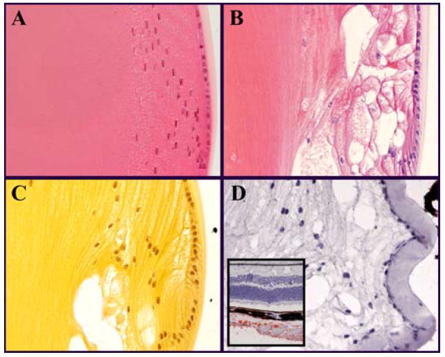
Figure 3.

Histological staining of the bow region at the equator of a 4-month-old lens. Sagittal section; anterior is oriented up. H&E stained sections of (A) WT and (B) SPARC-null lenses. Proteins stain pink and DNA stains violet. The large vacuoles in B appear to be empty and are surrounded by swollen cells. (C) Alcian blue with hematoxylin stained SPARC-null lens in which carbohydrates stain yellow. As with H&E alone, vacuoles do not stain. (D) Oil red O with hematoxylin stained lens in which lipids stain red. No lipid staining was observed in the lens while prominent red staining was observed in the sclera near the optic nerve entry (D, inset). Vacuoles at the SPARC-null lens periphery were not filled with proteins, carbohydrates, or lipids suggesting that abnormal fluid transport accounts for the cell swelling and vacuole formation in the absence of SPARC.
