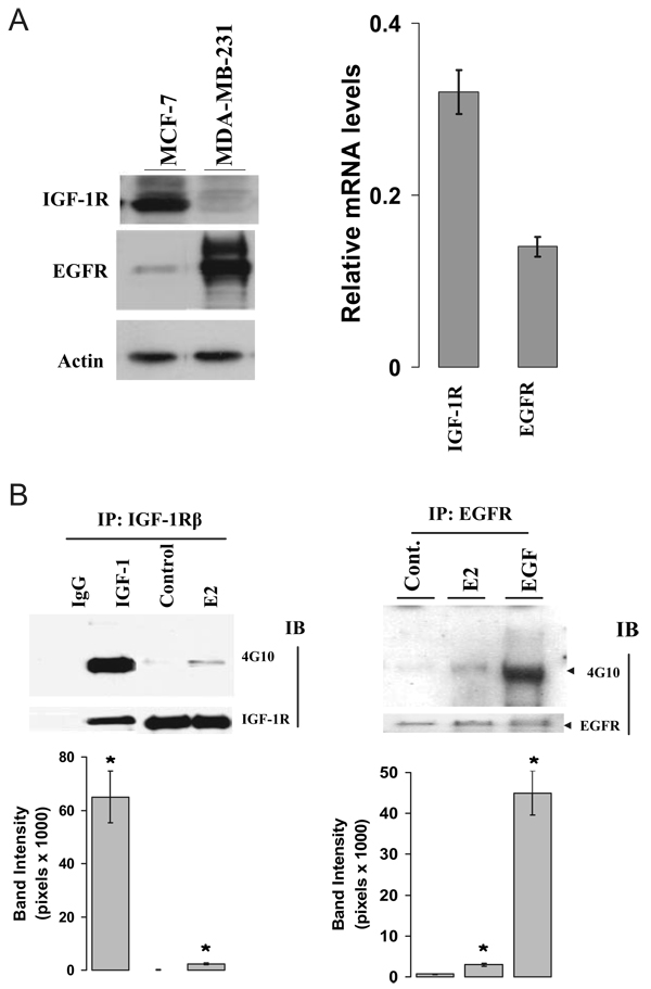
FIG. 2.
E2 activated both IGF-IR and EGFR in MCF-7 cells. A, Levels of IGF-IR and EGFR expression in MCF-7 cells. Both MCF-7 and MDA-MB-231 cells were extracted from cells at 80% confluence and processed for assessment of IGF-IR andEGFRexpression using Western blot (left) and RT-PCR method (right). B, Activation of IGF-IR and EGFR by E2. MCF-7 cells cultured in 1% DCC medium were stimulated with vehicle, 0.1 nM E2, 20 ng/ml IGF-I, or 20 ng/ml EGF for 5 min. Protein lysates were subjected to immunoprecipitation with anti-IGF-IR (left) or anti-EGFR (right) antibodies with subsequent immunoblotting using an anti-phosphotyrosine antibody (4G10) on Western blot. The nonspecific monoclonal IgG antibody was included in the immunoprecipitation step as a negative control. The membranes were further blotted with either anti-β-domain of IGF-IR or anti-EGFR antibodies for total receptor protein loading. All experiments were done at least three times. *, P < 0.05 compare with the vehicle-treated control.

