
Fig. 12.
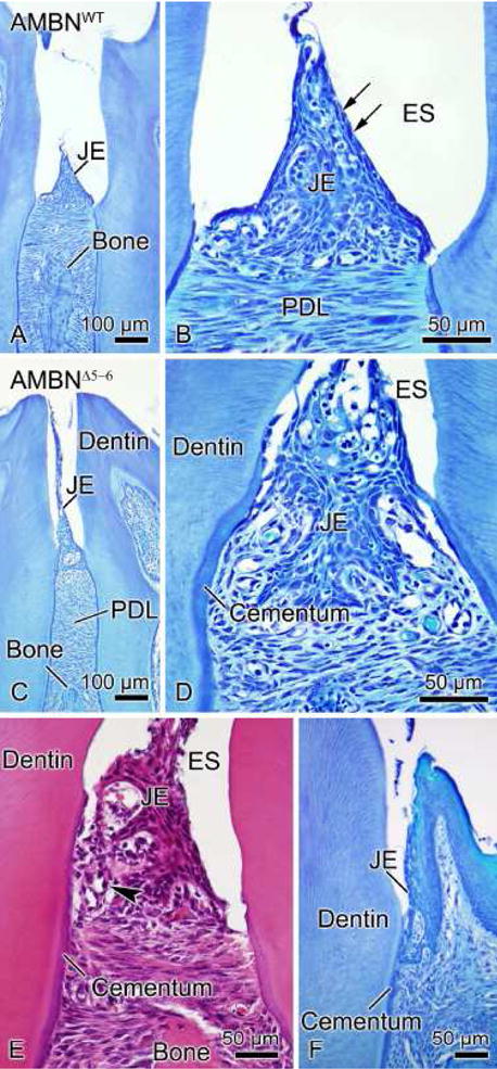
Light micrographs of the junctional epithelium (JE) in AMBNWT (A, B) and AMBNΔ5–6 mice (C–F). (A, B) The JE (arrows) is a stratified squamous non-keratinizing epithelium whose surface cells appear flattened and oriented parallel to the tooth surface. (C, D, E) Absence of expression of full-length AMBN gives rise to several structural defects in the interdental JE including detachment from the tooth surface and its repositioning onto cementum. The periodontal ligament (PDL) is also found lower on the root of the tooth. Inflammatory cells (arrowhead) infiltrated the JE of AMBNΔ5–6 mice. (F) Alterations in organization are less evident at the level of JE surrounding the labial and lingual aspects of molars. ES, enamel space.