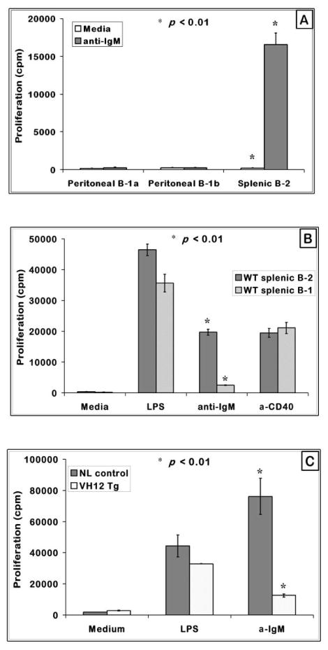
Figure 1. Peritoneal and splenic B-1 cells are hyporesponsive to BCR mediated proliferation.
(A) T cell-depleted splenic and FACS purified peritoneal B-1a and B-1b B cells from C57BL/6 mice were cultured at a cell density of 2.0 × 10⁵ cells/well with anti-IgM F (ab′)2 or anti-CD40 for 48 hours with indicated mitogens. Shown are the mean proliferation + SE from triplicate set of wells.
(B) FACS purified splenic B-1 (B220+ CD43+) and B-2 (B220+ CD43−) cells were plated at 2.5 × 10⁵ cells/well, treated with the stimulants at the indicated concentrations, cultured for 48 hours and measured for proliferative response by 3[H] Thymidine incorporation. The graph represents one of three independent assays.
(C) T cell-depleted VH12 transgenic splenic B-1 cells and negative control littermates were plated at a cell density of 2.5 × 10⁵ cells/well and cultured for 48 hours as in (B). Graph represents one of three independent assays. * indicates p < 0.01 when comparing B-1 and B-2 cell responses.

