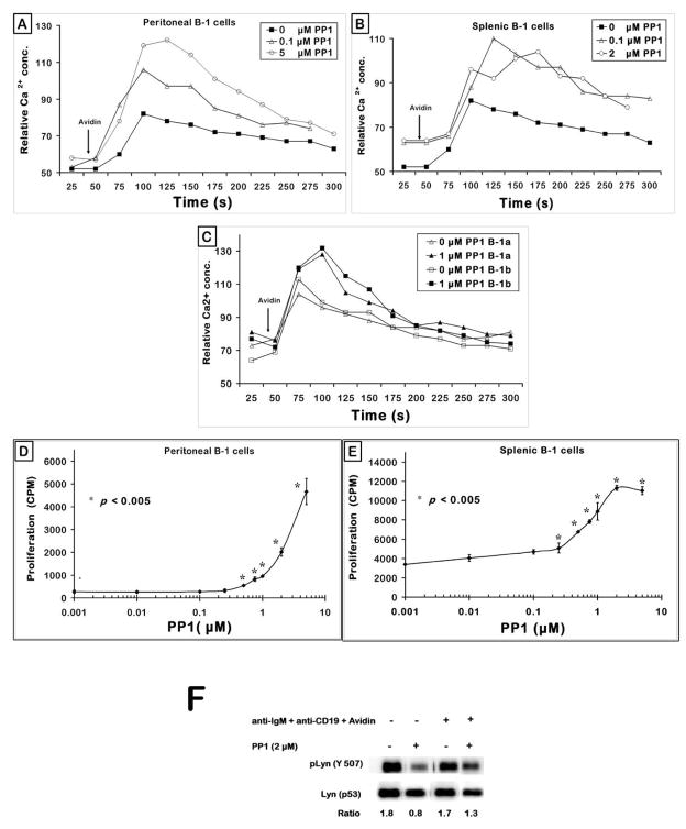
Figure 8. PP1 restores the BCR + CD19 signaling defect in B-1 cells.
(A) Peritoneal cells were loaded with Indo-1 and stained with α-B220 and α-Mac-1/CD11b as described in the Methods, treated with biotinylated anti-IgM and biotinylated anti-CD19; pre-incubated with PP1 at room temperature for 20 min prior to obtaining the 30 sec baseline. Avidin was added as indicated by the arrow. Graph represents one of three independent measurements.
(B) C57BL/6 splenocytes were Indo-1 loaded, stained with anti-B220~PE and anti-CD43~FITC, incubated with biotinylated anti-IgM F(ab′)2 and biotinylated anti-CD19, pre-treated with varying doses of PP1 for 20 min at room temperature, cell suspension warmed to 37°C, activated with avidin and calcium response monitored for 5 min. Shown were the responses of the gated splenic B-1 (B220+ CD43+) B cells at two doses of PP1 (μM).
(C) Cells from peritoneal lavage were treated as in (A), stained with α-B220~CY, α-Mac-1~PE, α-CD5~FITC, stimulated with biotinylated anti-IgM and biotinylated anti-CD19; pre-treated with PP1 at room temperature for 20 min prior to determining the 30 sec baseline calcium levels. Avidin was added as indicated by the arrow. Graph depicts the responses from gated B-1a (B220+ Mac-1+ CD5+) and B-1b (B220+ Mac-1+ CD5−) sub-populations.
(D) Peritoneal B-1 and VH12 transgenic splenic B cells (panel E) were treated with 50 μg/ml anti-IgM F (ab′)2 for 48 h in IF-12. The proliferative counts were determined as described in the Methods. Plot represents one of three independent in vitro proliferation assays. p < 0.005 compares the cpm at each of the PP1 doses to the 0 PP1 (μM) group i.e. anti-IgM alone group.
(F) Peritoneal B-1 cells were incubated with 2 μM PP1 for 30 min at 37°C. Cells were then treated with biotinylated antibodies to IgM and CD19 and stimulated with avidin for 10 min. The whole cell lysates were probed for pLyn (Y507), membrane was stripped and reprobed for total Lyn protein as loading control.

