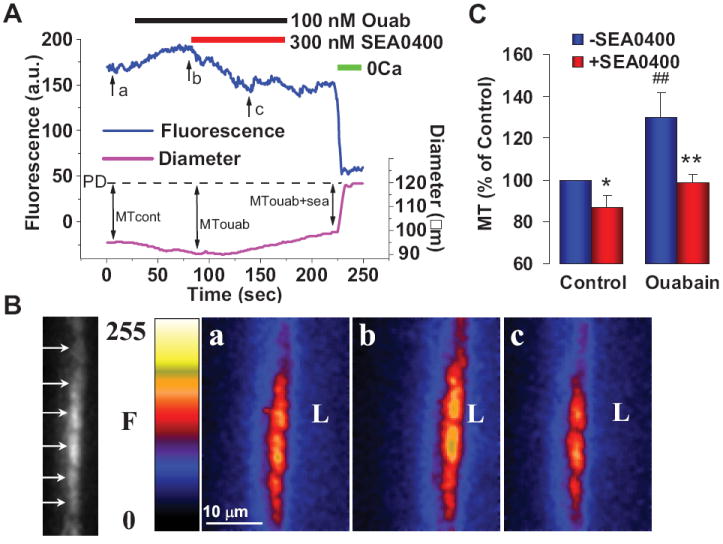
Figure 5.

Effects of low dose ouabain and SEA0400 on [Ca2+]CYT and myogenic tone (MT) in mouse pressurized mesenteric small arteries. A. Simultaneous recording of fluorescence (F, in arbitrary units, a.u., a measure of [Ca2+]CYT) and external diameter in a representative fluo-4 loaded normal mouse artery pressurized to 70 mm Hg. Bars at the top indicate periods of exposure to 100 nM ouabain, 300 nM SEA0400 and 0Ca medium (to determine passive diameter, PD). B. Arrows in the black and white spinning disk confocal image at the left indicate fluorescence in individual myocytes of a longitudinal cross-section through one wall of the artery in A. Pseudocolor images of this artery wall were captured at the times indicated by arrows “a” (control MT), “b” (MT with ouabain) and “c” (MT with ouabain + SEA0400) in A; “L” is located in the artery lumen. C. Summary of normalized MT data from this and five other, similar experiments. * = P < 0.05, ## = P < 0.01 vs untreated control; ** = P < 0.01 vs ouabain alone. Corrected from Ref. 66.
