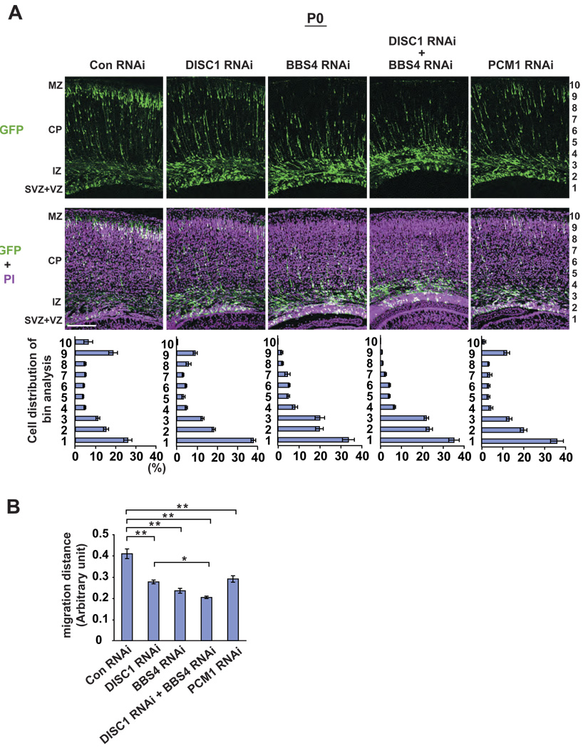
Figure 4. Knockdown of DISC1, BBS4, and PCM1 leads to neuronal migration defects in the developing cerebral cortex.
(A) RNAi constructs and GFP expression vectors were electroporated into the ventricular zone (VZ) at E15 and analyzed at P0. In brains with control RNAi (Con RNAi), 40% of GFP-labeled cells exited the VZ, and 25% of GFP-labeled cells completed migration and formed the superficial layers of the cortex that correspond to bins 9 and 10. By contrast, only less than 15% of GFP-positive cells reached the superficial layers in brain slices with DISC1 RNAi, BBS4 RNAi, or PCM1 RNAi, with the majority of GFP-positive cells remaining in the intermediate zone (IZ), subventricular zone (SVZ), and VZ. Green, cells co-transfected with GFP and RNAi constructs; purple, propidium iodide (PI). Scale bar: 100 µm.
(B) A migration distance is shown. Silencing of DISC1, BBS4, or PCM1 induces delayed radial migration (** P<0.0001). Silencing of both DISC1 and BBS4 expression leads to a more severe defect compared with that with either DISC1 RNAi or BBS4 RNAi. * P<0.05. Values are mean ± SEM.

