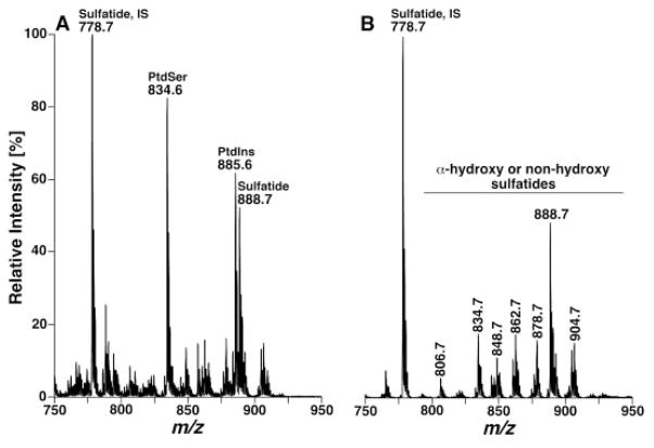
Figure 6.
Shotgun lipidomics analyses of sulfatide molecular species before and after treatment of a mouse cortex lipid extract with lithium methoxide in the negative-ion mode. The mass spectra in (A) and (B) were acquired directly from a lipid extract of mouse cortex before and after treatment with lithium methoxide, respectively, as illustrated in Fig. 3, by using a nanomate device. IS denotes internal standard. Both mass spectra are displayed after being normalized to the base peak in each spectrum.

