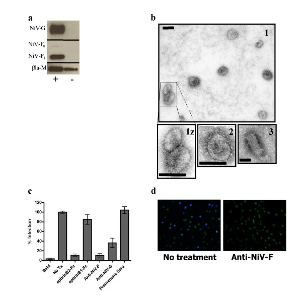
Figure 2.
βla-M+NiV-F/G VLPs morphologically, biochemically, and biologically mimic live NiV. a) VLPs produced in the presence (+) or absence (-) of envelope proteins were lysed and blotted for protein incorporation using anti-HA (NiV-G), anti-AU1 (NiV-F), or anti-NiV-M antibodies. b) Purified particles were analyzed under electron microscopy as described in materials and methods at 72,000× magnification. 1(z) = βla-M+NiV-F/G VLPs, 2 = NiV-M+F/G VLPs, 3 = pseudotyped VSV+NiV-F/G. Scale bars represent 100 nm. c) Vero cells were infected with NiV-F/G VLPs containing the βla-M fusion protein. Soluble ephrinB2-Fc and ephrinB1-Fc were added to a final concentration of 75 nM. Anti-NiV-F (834), anti-NiV-G (806), and pre-immune sera were added to a final concentration of 5 μg/ml. Infected cells (% blue positive) were quantified using flow cytometry with untreated entry (NoTx) normalized as 100%. Data shown as an average of triplicates from three individual experiments ± SEM. d) Fluorescence microscopy was performed on representative corresponding wells from (c) at 20× magnification using a beta-lactamase dual-wavelength filter (Chroma Technologies, Santa Fe Springs, CA).

