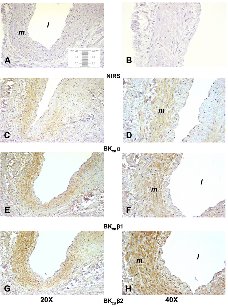
Figure 2.
Representative immunohistochemistry localizing BKCa subunits in the uterine artery from a nonpregnant woman. Panels A, C, E and G are at 20X magnification; panels B, D, F and H are at 40X. Panels A and B are controls with nonimmune serum, C and D are the pore α-subunit, E and F the β1-subunit, and G and H the β2-subunit. The vessel lumen is noted by the l and the media by m.

