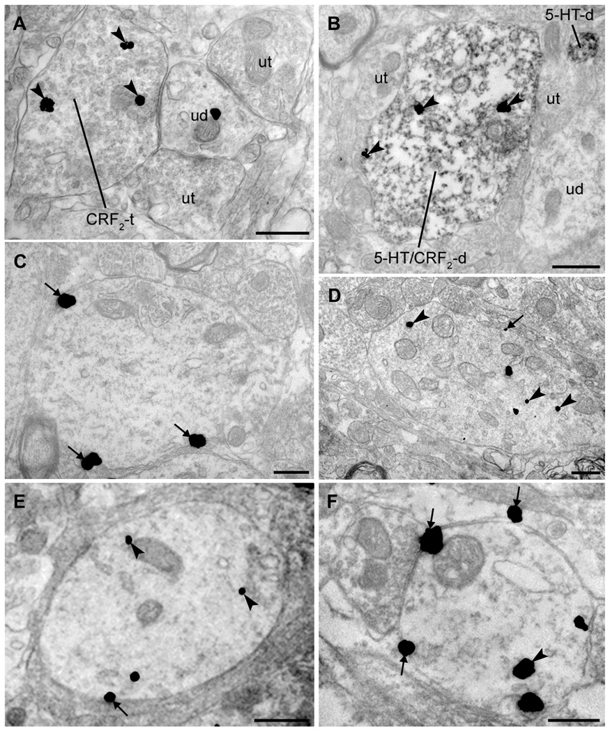
Figure 2. Ultrastructural examination of CRF receptors in the rat DR: distribution and effects of swim stress.
A: Some DR axon terminals contained evidence of CRF2 immunoreactivity (immunogold particles; arrowheads). CRF2-containing axon terminals (CRF2-t) were often found in synaptic contact with unlabeled dendrites (ud). Here, the ud targeted by the CRF2-t is also contacted by axon terminals lacking CRF2 immunoreactivity (ut). B: CRF2 is present in 5-HT-containing dendrites in the DR. Dual immunolabeling for CRF2 (immunogold; arrowheads) and 5-HT (immunoperoxidase) indicated that some 5-HT-containing dendrites colocalized CRF2 (5-HT/CRF2-d) in the DR. In this field, numerous unlabeled axon terminals (ut), a dendrite lacking detectable immunoreactivity (ud) and a dendrite containing 5-HT (5-HTd) but not CRF2 are also present. C–D: CRF1 and CRF2 have markedly different associations with the plasma membrane in unstressed rats. C: CRF1 was often found in association with the plasma membrane (arrows). D: In contrast to CRF1, CRF2 immunoreactivity was located predominantly in the cytoplasm of dendrites (arrowheads) although occasionally observed at the plasma membrane (arrows). E–F: Distribution of immunogold labeling for CRF1 and CRF2 in DR dendrites 24 h after swim stress. E: CRF1 is largely contained within the cytoplasm (arrowheads) with some receptor still present at the plasma membrane (arrows) following stress. F: CRF2 was redistributed to the plasma membrane (arrows) following swim stress, though some CRF2 immunoreactivity remained within the cytoplasm (arrowheads). Scale bars = 500 nm (A–F).

